


光顕用試料作製業務(受託業務)
Sample Preparations of Optical Microscope (Special service)
実験実習支援センターでは、光学顕微鏡で生物試料の微細構造を観察するための試料作製業務を請け負っています。
組織をパラフィン包埋し、薄切、染色するまでの行程をおこないます。
染色については、ヘマトキシリン・エオジン染色(HE染色)に限り、免疫染色等の特殊染色はおこないません。
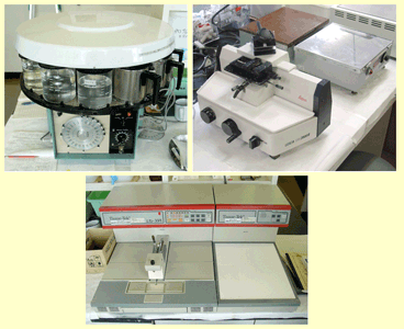
光顕用試料作製装置についてはこちらをご覧ください。
CRL provides services to prepare samples for observation of the microstructure of biological specimens with an optical microscope.
The process from paraffin-embedded tissue to thin sectioning and staining is performed.
Staining is limited to hematoxylin and eosin staining (HE staining), not immunostaining or other special staining.
Click here for more information about sample preparation equipment for optical microscopy.
業務内容 Contents
- パラフィンブロック作製 Paraffin block preparation・・・約27時間仕上がり Approx. 27 hours finish
自動包埋装置、エンベディングコンソールで脱水、包埋します
Dewatering with automatic tissue processor and embedding with tissue embedding console
標本は、採取後、固定した標本に限ります
Samples must be fixed after collection
- ブロック薄切 Thin sectioning・・・翌日仕上がり Next day finish
MASコートのスライドグラスに載せ、一昼夜伸展します
Paraffin sections are placed on MAS-coated glass slides and stretched overnight
- HE染色 HE staining・・・翌日仕上がり Next day finish
マイヤーのヘマトキシリンで3分〜30分、0.5%エオジンアルコールで1分〜5分染色
Staining with Meyer's hematoxylin for 3 to 30 minutes and 0.5% eosin alcohol for 1 to 5 minutes
光顕用試料作製の受付場所 Reception Location
4階 顕微鏡標本作製室1(405号室)
Microscopy Sample Preparation Room #1 (Room #405, CRL 4th floor)
担当者 Contact person
山元 武文(Takefumi Yamamoto)(TEL 2304)
福永 祥子(Sachiko Fukunaga)(TEL 2303)
岡本 久美(Kumi Okamoto)(TEL 2302)光顕用試料作製の依頼方法(学内向け)
How to request (SUMS only)
|
|
先頭へ TOP |
Last Updated 2023/09/01