


光学顕微鏡について Optical microscopes
光学顕微鏡には、鏡基の形式によって正立顕微鏡、倒立顕微鏡などと呼ばれ、また、光学系によっても位相差顕微鏡、微分干渉顕微鏡、蛍光顕微鏡などと区別されます。さらに、照明の仕組みから、明視野照明顕微鏡、暗視野照明顕微鏡、落射照明顕微鏡などがあります。
どのような試料の何を観察したいかによって適切な顕微鏡や観察方法を選ぶ必要があります。
Optical microscopes are classified as upright microscopes or inverted microscopes based on the configuration of the mirror base, and as phase contrast microscopes, differential interference contrast microscopes, or fluorescence microscopes based on their optical systems. Additionally, depending on the illumination system, there are bright-field microscopes, dark-field microscopes, and epi-illumination microscopes.
It is necessary to select the appropriate microscope and observation method based on the type of specimen and what you wish to observe.
光学顕微鏡の構造について Structure of optical microscope
光学顕微鏡は光学系の配置から、正立型と倒立型の二つに大きく分類され、それぞれはつぎのような構造をしています。
Optical microscopes are broadly classified into two types; upright and inverted based on the configuration of their optical systems. Each has the following structure.
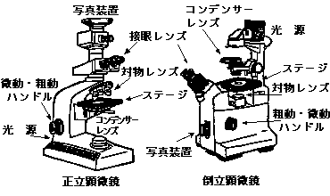
・正立顕微鏡 Upright microscope
この型の顕微鏡は、下部の光源からの照明光を反射鏡、視野絞り、コンデンサーレンズを通過させ試料にあてます。ステージの上方に対物レンズがあり、ステージと対物レンズの間に試料を載せ、上部の接眼レンズから試料を観察します。また、写真撮影装置は上部にあります。
An upright microscope is a standard type of microscope in which the objective lens is positioned above the stage, allowing the specimen to be observed from above. Light from a light source at the bottom passes through the reflector, the field diaphragm and the condenser lens before illuminating the specimen. The microscope is fitted with an accessory for photography at the top.
・倒立顕微鏡 Inverted microscope
この型の顕微鏡は、照明装置が上部にあり、照明光はコンデンサーレンズ、視野絞りを通ってステージの上の試料にあたります。ステージの下に対物レンズがあり、結像光線を下部の反射鏡で反射させ、接眼レンズで観察します。従って、培養細胞などの入ったシャーレなどをステージの上に置いた場合でも、観察する面がシャーレの底面になり、スライドグラスを用いたときと同様に明瞭に観察することが出来ます。
そして、倒立顕微鏡、正立顕微鏡はいずれの顕微鏡も適当な付属装置を付けることによって、位相差顕微鏡や微分干渉顕微鏡、あるいは蛍光顕微鏡として使用出来ます。
An inverted microscope is a microscope in which the objective lens is positioned below the stage, allowing the specimen to be observed from below. The illumination system is located at the top, and the light passes through the condenser lens and the field diaphragm to illuminate the specimen on the stage. Because it provides ample space on the stage, it is ideal for observing living cells in culture medium (live-cell observation), conducting experiments using petri dishes or flasks, and inspecting large samples such as metals.
Both inverted and upright microscopes can be used as phase contrast microscopes, differential interference contrast microscopes, or fluorescence microscopes by attaching appropriate accessories.
対物レンズ Objective lens
光学顕微鏡の分解能は、光源の波長のほかに、対物レンズの開口数と収差によって決まります。従って、対物レンズの性能に因って大きく左右されます。そして、対物レンズの開口数は、レンズの倍率に関係があり、収差は、レンズによる光の屈折に関係があります。そこで、対物レンズの性能を上げるために、収差を取り除いたり、開口数を大きくするなど様々な工夫が施されています。
The resolution of an optical microscope is determined by the wavelength of the light source as well as the aperture and aberration of the objective lens. Consequently, it is largely dependent on the performance of the objective lens. The numerical aperture of the objective lens is related to the lens magnification, while aberrations are related to the refraction of light by the lens. Therefore, various measures are taken to improve the performance of the objective lens, such as correcting aberrations and increasing the numerical aperture.
・収差について Aberration
収差には、球面収差、コマ収差、非点収差、色収差、像面湾曲収差などがありますが、中でも対物レンズの分解能に大きな障害を与えるのが色収差と像面湾曲収差の二つです。そこで、この二つを補正するために、凸レンズと凹レンズを組み合わせた様々な構造の対物レンズがあります。たとえば、アクロマート対物レンズ:このレンズは、C線(赤)とF線(青)の色収差を完全に補正しています。
アポクロマート対物レンズ:これは、C線、F線とD線(黄)の色収差を補正しています。
セミアポクロマート対物レンズ:このレンズは、アポクロマート対物レンズの設計をやや簡単にしたものです。
プランアクロマート対物レンズ:このレンズは、C線とF線についての色収差と、像面湾曲収差について補正したもので、写真撮影に適しています。
プランアポクロマート対物レンズ:このレンズは、C線、F線とD線の色収差と像面湾曲収差について補正したもので、収差補正に関してはほぼ完璧な対物レンズです。
Aberrations include spherical, coma, astigmatism, chromatic and field curvature aberration; among these, chromatic and field aberration greatly impair the objective resolution. Therefore, to correct these two aberrations, objective lenses with various configurations combining convex and concave lenses are used. For example, achromatic objective lenses completely correct chromatic aberration in the C-line (red) and F-line (blue).
Apochromat: a lens that corrects chromatic aberrations in the C (red), F (blue), and D (yellow) lines.
Semi-apochromat: a lens that is a slightly simplified version of the apochromat lens design.
Plan-achromat: a photographic lens that corrects chromatic aberrations in the C (red) and F (blue), and field curvature aberration.
Plan-apochromat: a lens that corrects chromatic aberrations in the C (red), F (blue) and D (yellow) lines, and field curvature aberration. It offers virtually perfect correction of aberrations.
・開口数とは? Numerical aperture
開口数とは、顕微鏡の対物レンズや光ファイバーなどが、どれだけ広い角度の光を集められるかを示す指標です。
識別できる2点の最小距離を分解能といい、これは 0.61×λ/n sinθ(θ:光源とレンズの光軸のなす角、n:レンズと試料の間の媒質の屈折率、λ:光源の波長)という式で表され、この式の n sinθをレンズの開口数と呼びます。開口数が大きいほど分解能が上がりますが、レンズの構造上θは72度が限界です。「油浸対物レンズ」は、カバーグラスと対物レンズの間をガラスと同じ屈折率を持つシーダ油(n = 1.515)で満たすことによって開口数を大きくし、分解能を上げようとするものです。
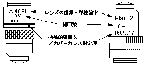
対物レンズの表示は、図に示すように、倍率、開口数、用途、カバーガラス厚等が付けられています。
なお、対物レンズには、図に示します様に色々な記号が表示されています。これらはそれぞれ図に示した意味を持っております。
the numerical aperture (NA) is a dimensionless number that characterizes the range of angles over which the system can accept or emit light.
The resolution is based on the minimum distance at which the points can be distinguished as individuals, which is expressed by the formula 0.61×λ/n sin θ (θ: the angle between the light source and the optical axis of the lens, n: the index of refraction of the medium in which the lens is working, λ: the wavelength of the light source); in this formula, “n sin θ” is called the NA of the lens. Although a larger NA results in higher resolution, the lens design limits θ to 72 degrees. Oil immersion objective lenses are designed to increase the NA and improve resolution by filling the space between the cover glass and the objective lens with cedar oil (n = 1.515), which has the same refractive index as glass.
As shown in the figure, the objective lenses are labeled with their magnification, NA, intended use, cover glass thickness, and other specifications.
接眼レンズ Eyepieces
接眼レンズは、1〜3個のレンズを組み合わせて構成されており、鏡筒内に付属している視野絞りの位置でホイゲンス型とラムスデン型の2種類に分ける事が出来ます。ホイゲンス接眼レンズは、視野絞りが接眼レンズの鏡筒の中にあり、一方、ラムスデン接眼レンズでは、この視野絞りが接眼レンズの鏡筒の外にあります。
また、ペリプラン接眼レンズと呼ばれるものは、非点収差、像面湾曲収差、倍率色収差を補正し、視野の周辺部まで鮮明に見える様にしたもので写真撮影に適しています。
Eyepieces consist of a combination of one to three lenses and are classified into two types based on the position of the visual field diaphragm inside the tube: Huygens and Ramsden eyepieces. In a Huygens eyepiece, the field diaphragm is located inside the tube, whereas in a Ramsden eyepiece, the field diaphragm is located outside the tube.
In addition, Periplan eyepieces correct for astigmatism, field curvature, and chromatic aberration, ensuring a sharp image all the way to the edges of the field of view, making them suitable for photography.
・視野絞りとは? Field diaphragm
これには、接眼レンズ鏡筒内で発生する有害な反射光や周辺部の収差の多い部分をカットする働きがあります。視野絞りは、接眼レンズの焦点面にありますので、この位置に計測用のミクロメーターを置きますと、試料の観察と同時にミクロメーターの目盛りで試料の大きさの計測も出来ます。
The field diaphragm serves to block harmful reflected light generated inside the eyepiece tube and areas with high aberration at the periphery. Since it is located at the focal plane of the eyepiece, placing a measuring micrometer at this position allows you to measure its size using the micrometer’s scale as well as to observe the specimen.
位相差顕微鏡 Phase contrast microscope
無色透明な媒質の中に、媒質とごく僅かだけ屈折率が異なる無色透明な微細構造
がある場合、原則として、光学顕微鏡でこれを観察することは出来ません。これを解決するために染色を行ないます。しかし、染色せずに肉眼で観察しようとして開発されたのが位相差顕微鏡です。
With a standard optical microscope, light passes right through transparent cells, making them impossible to observe due to a lack of contrast. Staining is typically performed to resolve this issue, but the phase-contrast microscope made it possible to quantify cellular structure and components without using fluorescence.
・なぜ見えるようになるか Working principle
位相差顕微鏡は、極くわずかな屈折率の差を明暗の差に換えて観察するように工夫された顕微鏡です。
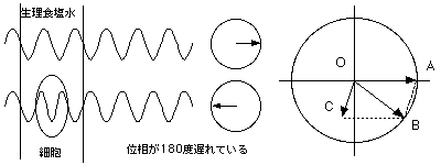
いま、生理食塩水に浮遊した細胞を観察するとします。細胞は周囲の媒質である生理食塩水より屈折率が大きいので、細胞を通過した光は、生理食塩水を通過した光より光学的光路長がごく僅か長くなります。光学的光路長とは、媒質の幾何学的な厚さにその媒質の屈折率をかけた値です。下図に示しますように、生理食塩水より屈折率の大きい細胞を通過した光OBは、バックグラウンドである生理食塩水を通過した光OAに比べ僅かながら位相の遅れが生じます。
ところで、光OBは、生理食塩水を通過した光OAと細胞で回折を起こした光OCとによって合成された光です。光OCは、光OAより振幅が小さく、位相が1/4波長以上遅れています。そこで、位相差板を用いて、像面における回折光線成分の光OCの位相を、非回折光線成分の光OAより1/2波長だけ遅らせてやると、両者の合成波である光OBの振幅は(OA−OC)となり、細胞の像は、バックグラウンドである生理食塩水の明るさよりも暗くなって見える筈です。反対に、非回折光線成分の光OAの位相を位相差板で遅らせて、回折光線成分の光OCと同じ位相にしてやると、両者の合成波の光OBの振幅は(OA+OC)となり、細胞の像はバックグラウンドである生理食塩水の明るさより明るくなって見える筈です。このように位相差顕微鏡は、僅かな屈折率の差を利用して我々の目には見えなかった微細構造を見えるようにしたものです。
A phase contrast microscope is an optical microscope that converts phase shifts in light passing through a transparent specimen to brightness changes in the image.
When observing cells suspended in saline solution, because the cells have a higher refractive index than the surrounding medium, saline solution, the optical path length of light passing through the cells is slightly longer than that of light passing through the saline solution. Optical Path Length refers to the product of the physical path length that light travels through a medium and the refractive index of that medium. As shown in the figure below, the light OB passing through a cell, which has a higher refractive index than the saline solution, is slightly phase-shifted relative to the light OA passing through the background saline solution.
The light OB is synthesized by the light OA, which has passed through saline solution, and the light OC, which has diffracted within the cell. OC has a smaller amplitude and is phase-shifted by -90°relative to OA. When the diffracted light OC is phase-shifted by -180° by passing it through the phase shifter relative to the non-diffracted light OA, the amplitude of the combined wave OB will be “OA-OC”, resulting a darker foreground (cells) and a lighter background (saline solution). While, if OA is phase-shifted by -180°by the phase shifter so that it matches the phase of the diffracted light component OC, the amplitude of OB will be “OA+OC”, resulting a lighter foreground (cells) and a darker background (saline solution). In this way, phase-contrast microscopy is an optical microscopy technique that converts phase shifts in light passing through a transparent specimen to brightness changes in the image. Phase shifts themselves are invisible, but become visible when shown as brightness variations.
・位相差顕微鏡を使うとき対物レンズに対応した絞りを用いる Using the appropriate diaphragm for the objective lens
位相差顕微鏡では、絞りはドーナツ型のリングスリットを採用しており、コンデンサーと一体になっています(ターレットコンデンサー)。対物レンズの倍率が決まると、それに対応した絞りの輪の直径と幅が決まりますので、各対物レンズに対応した絞りを用いる必要があり、対物レンズを交換する度に絞りの方も取り換えなければなりません。
位相差対物レンズには、屈折率の大きい観察対象を周囲の媒質より暗くする、ポジティブコントラスト(ダークココントラスト)対物レンズと、これとは逆に、屈折率の大きいものをバックグラウンドより明るくするネガティブコントラスト(ブライトコントラスト)対物レンズとがあります。
Phase-contrast microscopes are equipped with a doughnut-shaped ring slit diaphragm integrated with a condenser (turret condenser). Once the magnification of the objective lens is determined, the diameter and width of the diaphragm are also determined; therefore, it is necessary to use a diaphragm that matches the objective lens, and the diaphragm must be replaced each time the objective lens is changed.
There are two types of phase contrast objectives: positive-contrast (dark-contrast) objectives, which have a high refractive index and make the observed object appear darker than the surrounding medium; and negative-contrast (bright-contrast) objectives, which have a high refractive index and make the observed object appear brighter than the background.
微分干渉顕微鏡 Differential interference contrast microscope
微分干渉顕微鏡は、光学顕微鏡に微分干渉装置を取付けて、光の干渉現象を利用して、試料の屈折率や厚みの変化を干渉色の変化や明暗のコントラストに換えて観察出来る様にしたもので、明視野顕微鏡では観察出来ない透明な試料、例えば無染色の生物試料などを観察するのに用います。
A differential interference contrast microscope is an optical microscope equipped with a differential interference contrast (DIC) unit, and works on the principle of interferometry. It converts changes in the refractive index or thickness of the sample into variations in interference colors and brightness contrast, thereby enabling observation of transparent samples, such as unstained biological specimens that cannot be observed using a brightfield microscope.
・微分干渉装置について Differential interference contrast (DIC) unit
微分干渉装置は、一組のノマルスキープリズムとポラライザー、アナライザーと呼ばれる二枚の偏光板から出来ています。これを光学顕微鏡に下図の様な配置で取り付けることによって微分干渉顕微鏡として使用することが出来ます。
A DIC unit consists of a set of Nomarski prisms and two polarizing filters, known as a polarizer and an analyzer. By mounting this unit on an optical microscope, it can be used as a differential interference microscope.
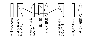
微分干渉装置により、光源からの光はポラライザを通過して直線偏光に変換され、コンデンサー側ノマルスキープリズムに入射し、偏光面が互いに直交した2つの直線偏光に分かれます。2つに分離した直線偏光が再び交わる面に焦点面を一致させてコンデンサーレンズを置くと、わずかに横ずれした互いに平行な光が得られます。この平行光線を試料に照射します。
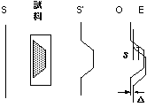
ここで、媒質内に屈折率の異なる透明物体がある試料を考えてみます。
明視野で照明した場合、図の様に入射波面SはS'のように変形されて出てきますが、先の2つの平面偏光で照明すると、図の様に出てきた光は僅かに横ずれした2つの波面、OとEになります。
試料を透過した光は、対物レンズを通り焦点を結びますが、この焦点の位置に対物側ノマルスキープリズムのローカライズ面を一致させておくと、2つの光路は再び1つになり2つの波面が干渉します。この時、波面に傾斜のある部分の光路差はδとなり、背景の光路差Δとは異なるために、それぞれ異なった干渉色を示します。この干渉色を観察することにより、波面の傾斜部分、つまり屈折率の変化する部分を見ることができます。また、背景の光路差Δは、対物側ノマルスキープリズムを移動することにより変化させることができます。
DIC works by separating a polarized light source into two orthogonally polarized mutually coherent parts which are spatially displaced (sheared) at the sample plane, and recombined before observation. Light emitted from the source enters the polarizer and is polarized at 45°. The polarized light enters the first Nomarski prism and is separated into two rays polarized at 90° to each other. The two rays are focused by the condenser for passage through the sample. These two rays are, however, not quite aligned, with one lying slightly offset with respect to the other.
Suppose we have a sample containing a transparent object with different refractive indexes in the medium.
When illuminated with bright-field illumination, the incident wavefront S is distorted as S', as shown in the figure; however, when illuminated with the two plane-polarized lights mentioned earlier, the emerging light consists of two wavefronts, O and E, which are slightly offset from each other, as shown in the figure.
The lights travel through the objective lens and are focused for the second Nomarski prism. The second prism recombines the two lights into one polarized light, and the combination of the lights leads to interference. At this point, the path difference in the inclined portion of the wavefront is δ. Since this differs from the path difference Δ in the background, they produce different interference colors. By observing these interference colors, we can identify the inclined portion of the wavefront, that is, the region where the refractive index changes. Furthermore, the path difference Δ in the background can be adjusted by moving the objective Nomarski prism.
・微分干渉顕微鏡を用いて観察するには Observation using a differential interference contrast microscope
先ず、ノマルスキープリズムとポラライザー、アナライザーを光路からはずし、通常の顕微鏡と同様に、コンデンサーレンズの芯出しを行います。
次に、ポラライザーとアナライザー(対物側ノマルスキープリズムが一体になっているものもある)を光路に入れ、ポラライザー回転つまみを操作しポラライザーがアナライザーと直交する位置に固定します。この位置決めをするには、接眼レンズをはずし、かわりに芯出し鏡を取り付けて覗いた時の視野に見える暗十字、または黒の干渉縞(機種によって異なる)が、最もシャープに見える位置に合わせます。
最後に、ターレットを回すことにより対物レンズの倍率に応じたコンデンサー側ノマルスキープリズムを選択し、対物側ノマルスキープリズムを光路に入れて観察します。
また、観察中に移動つまみを用いて対物側ノマルスキープリズムをずらしますと、背景の干渉色が連続して変化します。
First, remove the Nomarski prism, polarizer, and analyzer from the optical path, and center the condenser lens as with a standard microscope.
Next, insert the polarizer and analyzer (some models have an integrated objective Nomarski prism) into the optical path, and use the polarizer rotation knob to fix the polarizer in a position perpendicular to the analyzer. To perform this alignment, remove the eyepiece and replace it with an alignment mirror. Then, adjust the position so that the dark cross or black interference fringes (which vary by model) visible in the field of view appear as sharp as possible.
Finally, rotate the turret to select the condenser Nomarski prism corresponding to the objective lens magnification, and place the objective Nomarski prism in the optical path to begin observation.
Additionally, if you adjust the position of the objective Nomarski prism using the adjustment knob during observation, the interference color of the background will change continuously.
蛍光顕微鏡 Fluorescence microscope
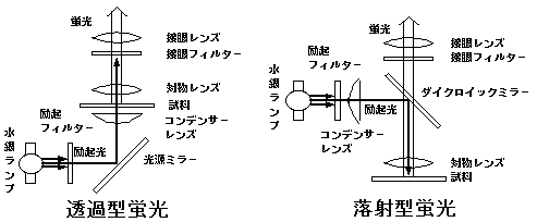
蛍光顕微鏡は、試料に紫外線や短波長の可視光線(紫色〜青色)を照射し、励起された試料より発する蛍光を観察するために、光学顕微鏡に、試料を励起するための光源部と、励起光をカットし、試料より発した蛍光を透過させる接眼フィルターを取り付けた装置です。透過型蛍光顕微鏡(薄い試料の観察に用いる)と落射型蛍光顕微鏡(厚い不透明な試料の観察に適する)の2つがあります。落射型蛍光顕微鏡では、短波長の光を反射し長波長の光のみ透過させる性質を持ったダイクロイックミラーを光路に挿入し、光源からの励起光と試料からの蛍光を分離しています。
A fluorescence microscope is an optical microscope equipped with a light source to excite the sample and an eyepiece filter that blocks the excitation light while allowing the fluorescence emitted by the sample to pass through. It works by illuminating the sample with ultraviolet light or short-wavelength visible light (purple to blue) and observing the fluorescence emitted by the excited sample. There are two types of fluorescence microscopes: transmission fluorescence microscopes, which are used for observing thin specimens, and epifluorescence microscopes, which are suitable for observing thick, opaque specimens. In the latter, a dichroic mirror, which reflects short wavelength light and transmits only long wavelength light, is inserted into the optical path to separate the excitation light from the light source from the fluorescence emitted by the sample.
・フィルター Filter
光源としては、一般的には、超高圧水銀ランプが用いられています。この光源は365 nm、405 nm、436 nmに輝線を持っています。
ところで、この光源からは、種々の波長の可視光線も放射されており、観察の妨げとなりますので、励起フィルターを用いて、紫外線と短波長の可視光線のみを透過させ、不要の可視光線を吸収除去しています。
また、試料から発した蛍光を観察する際に観察者の目を紫外線から保護する為に接眼フィルター(バリアフィルター)を用います。このフィルターは、蛍光によって起こる霧視現象を防止し試料の蛍光を明瞭に観察出来る様にする為のものでもあり、蛍光顕微鏡で最も重要な部分です。
The light source typically consists of an ultra-high-pressure mercury lamp, which has emission lines at 365 nm, 405 nm, and 436 nm. Since the light source also emits visible light of various wavelengths, which can interfere with observation, an excitation filter is used to transmit only ultraviolet and short wavelength visible light, while absorbing and removing unwanted visible light.
In addition, an eyepiece filter (barrier filter) is used to protect the observer’s eyes from ultraviolet light when observing fluorescence emitted by the sample. This filter also prevents the blurring of vision caused by fluorescence, allowing for clear observation of the fluorescence from the sample, and is the most critical component of a fluorescence microscope.
・蛍光顕微鏡による試料の観察法 Observing samples using fluorescence microscopy
試料そのものが発蛍光性である場合と、試料に処理を施して蛍光性に換えて観察する場合とがあります。後者には、蛍光染色法、化学的蛍光処理法、蛍光抗体法等があります。
蛍光染色法は、試料を蛍光色素で染色する方法です。次の、化学的蛍光処理法は、試料に化学的処理を施して蛍光性をもたせる方法で、サイアミンを酸化してチオクロームに変えて青白色の蛍光を観察したり、ホルムアルデヒドを作用させてモノアミン類を観察したりするのがこれに相当します。最後の蛍光抗体法は、免疫組織化学的方法で、細胞や組織内の抗原の存在分布を検索する方法として、細胞生物学、微生物学、免疫学、病理学、臨床検査などの諸分野で広く用いられている方法です。抗原性を有するもので、これに対する蛍光標識抗体があれば、この方法による観察の対象になります。
There are two main approaches: observing samples that are inherently fluorescent and observing samples that have been treated to make them fluorescent. The latter includes fluorescent staining, chemical treatment with fluorescent, and fluorescent labeled antibody.
Fluorescent staining involves staining the sample with a fluorescent dye, while chemical fluorescent treatment is a method of imparting fluorescence to samples through chemical processing. The fluorescent antibody method is a type of immunohistochemical technique widely used in various fields, including cell biology, microbiology, immunology, pathology, and clinical testing, to detect the presence and distribution of antigens within cells and tissues. Any tissue that possesses antigenicity and for which a fluorescently labeled antibody is available can be observed using this method.
|
|
先頭へ |
Last Updated 2005/06/22