


多重蛍光免疫組織化学法
〜共焦点レーザー走査顕微鏡でこんなに簡単、でも....〜
Immunohistochemistry has developed into a standard morphological method in life science research.
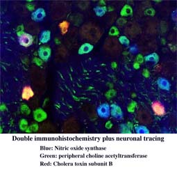
Among techniques used in immunohistochemistry, multiple immunohistochemistry using two or more
antibodies has become extremely convenient and popular because of recent improvements in fluorescent
markers and microscopes.
However, this popularization may be generating a lot of incorrect and insufficient data which can be seen even in published papers.
This lecture and wet-lab practice will provide you not only with how to do "easy" multi-fluorescent immunohistochemistry but also "practical tips" in immunohistochemistry to avoid such mistakes.
免疫組織化学法は生命科学分野における形態研究法の標準手法となっている。様々な免疫組織化学法のなかでも、複数の抗体を用いる多重免疫染色法は、蛍光標識や顕微鏡の進歩・改良により飛躍的に簡便になり、誰もが使えるように一般化した。しかし反面、このような一般化によって、誤りや不完全な結果が雑誌に掲載されているような論文の中にすら見られるようになっている。
本講義・演習では、「簡単」になった多重免疫組織化学染色法だけでなく、かような過ちを回避する「実践的なポイント」を学んでいただきたい。
|
|
先頭へ |
Last Updated 2005/6/22